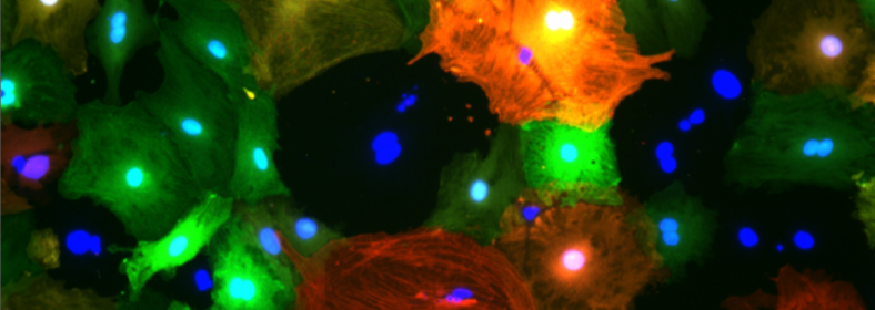
The iconic #TRAINFORINNOVATION image was taken using a fluorescence microscope at the ICGEB Trieste laboratories and shows murine cardiac fibroblasts (which are cells that make up our connective tissue). The fibroblasts express Red Fluorescent Protein (RFP) under an alpha smooth muscle actin promoter, and Green Fluorescent Protein (GFP) under a collagen promoter. This means that whenever the cells are producing smooth muscle actin, they also produce RFP, and whenever they are producing collagen, they also produce GFP.
Fibrosis is a thickening and scarring of connective tissue, for example after a heart attack, severely impairing functionality of the heart. During fibrosis, fibroblasts differentiate into myofibroblasts. The RFP GFP fibroblasts used in these experiments can act as a model for fibrosis, as they differentiate into myofibroblasts in culture. When they are differentiated into myofibroblasts, they express higher amounts of smooth muscle actin and collagen, which means the cells can be seen to be more red and more green under a fluorescent microscope. In order to find chemical compounds which can be used to combat fibrosis, these cells are treated with various compounds, to see which compounds will block a differentiation into myofibroblasts and therefore reduce the production of RFP and GFP.
In order to process the large amounts of data produced in this study, and to find more interesting compounds, the researchers at the Jozef Stefan Institute in Ljubljana are using specialised machine learning algorithms. Together, the researchers are working to identify new compounds that can be used to treat fibrosis.
Operation co-funded by the European Regional Development Fund